
Breast Augmentation
Your options, your operation and your long-term care.
01 · Understand your choices
The basics & your options
Breast augmentation may increase size, fullness and projection, restore volume after pregnancy or weight loss, and address asymmetry or developmental shape differences.
Choose surgery for yourself. Improved self-image is a personal hope, not a guaranteed effect on confidence, relationships or body silhouette. Implants do not correct severe drooping: a lift alone or a lift with implants may be appropriate, at the same operation or in stages.
Filling, shape and surface are different choices

Filling (left) and shape (right) are separate features; the drawings do not show available models.
Saline or silicone gel
Saline: a silicone shell contains sterile salt water. A leak usually causes deflation; the fluid is absorbed by the body, but the implant still needs assessment.
Silicone gel: the feel may be closer to breast tissue. Rupture can be silent without obvious collapse; examination alone cannot rule it out. Gel may remain in the capsule or escape beyond it. Ultrasound or MRI is used for assessment.
Consistency, shape & profile
More cohesive silicone gel is more form-stable, but not rupture-proof. Gel consistency and shape are separate: cohesive implants can be round or anatomical. Some firmer devices may need a longer incision.
Round implants may add upper fullness; higher profiles increase projection for a given base width. Rotation has less effect on round shape, but displacement is still possible. Anatomical rotation can distort breast shape.
Surface: smooth implants can move in the pocket and may ripple. Textured surfaces interact differently with scar tissue, but are not simply “safer” or a blanket way to prevent contracture. Textured implants have a higher BIA-ALCL risk; use depends on the specific product and local approval.
Fat transfer for selected, modest augmentation
Your own fat is removed by liposuction, processed and reinjected into the breasts. Adequate donor fat is needed. Some fat is reabsorbed, so retained volume varies and repeat treatment may be needed. Risks include cysts or oil cysts, calcifications, fat necrosis, infection and imaging changes; tell your radiology team. A breast expansion system is not routinely required.
Not all options are offered or available. Discuss the exact manufacturer, model, size, patient leaflet and device card with your surgeon. Eligibility depends on maturity, local regulations and the specific device.
02 · Before surgery
Consultation & preparation
A good consultation connects your goals with your anatomy, health and willingness to accept long-term follow-up. Bring your questions; uncertainty or anxiety deserves discussion.
Could surgery be right for you?
- Good general health, fully developed breasts and realistic expectations.
- Concern about small size, upper-breast emptiness, loss of volume or shape after pregnancy, weight change or aging.
- Asymmetry, elongated shape or incomplete development of one or both breasts.
Discuss pregnancy and breastfeeding plans and suitable timing. Breastfeeding after surgery cannot be guaranteed. Choosing no surgery is always an option.
Your health history
- Goals, desired size and shape; medical conditions, allergies and previous treatments or surgery.
- All medicines, vitamins and herbal products; alcohol, tobacco and other drug use.
- Family breast-cancer history; prior breast imaging, biopsies, pregnancy and breastfeeding.
What the qualified surgical team will do
Review your health and risk factors; examine and measure breast size, shape, skin quality and nipple/areola position; and take clinical photographs with consent. Discuss implants, fat transfer, a lift or no treatment, pocket and incision choices, anesthesia, likely outcomes and limitations, risks and possible revision.
Prepare safely
- Tests if indicated
- Review medicines
- Stop smoking as advised
- Review bleeding risks
- Ride & adult support
Laboratory tests or breast imaging are ordered when indicated by your health, examination and age/risk-based screening guidance; a mammogram is not mandatory at every age. Follow individualized smoking-cessation advice.
Review aspirin, anti-inflammatory medicines and herbal supplements because some increase bleeding risk. Do not stop or change prescribed medicines without instructions from your treating team. Ask about fasting and which medicines to take on the day.
Plan your return home
Surgery should take place in an appropriately approved, equipped facility with a qualified anesthesia team. After intravenous sedation or general anesthesia, arrange a ride and a responsible adult for the first night. Discharge follows an individual recovery assessment, not a promised number of hours.
Before consent, ask why the proposed device and technique suit you, what alternatives exist, and who will help if a problem occurs. Receive and discuss the manufacturer’s patient information.
03 · Your individualized plan
The procedure, step by step
Implant surgery is tailored to your anatomy, tissue coverage, activity, goals and chosen device. Fat-transfer steps differ and will be explained if selected.
Anesthesia
Intravenous sedation or general anesthesia may be used. Your surgical and anesthesia teams recommend the appropriate approach for you and the facility.
Incision
Options include the lower semicircle at the areolar border, a curved incision in the fold beneath the breast, or a short incision in an armpit crease. Choice depends on anatomy, implant, enlargement and the shared surgical plan.
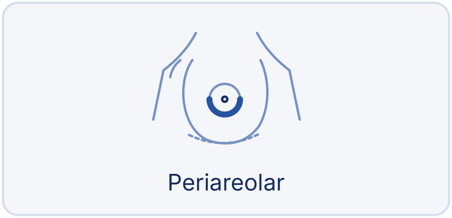
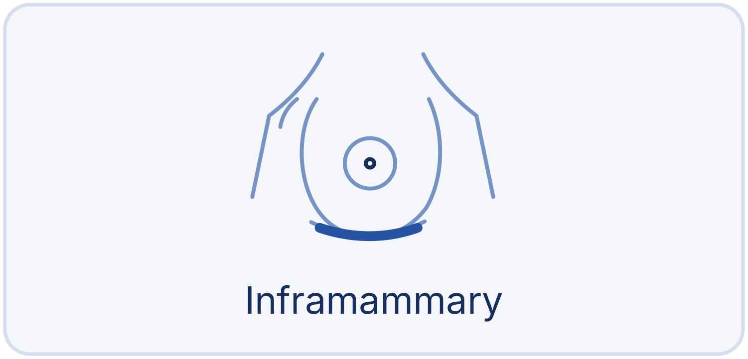
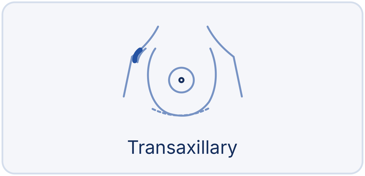
Front-view maps. Bold blue marks the incision; the pale dashed line marks the breast fold. No breast-lift scar pattern is shown.
Pocket and implant placement
The pocket may be behind the gland and over pectoralis, or under/partly under pectoralis, including dual-plane techniques. Not every “under-muscle” implant is fully covered by muscle. Coverage, movement and appearance trade-offs are individualized.
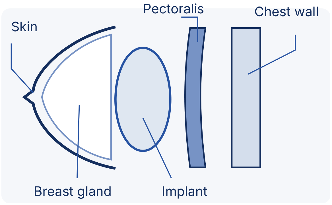
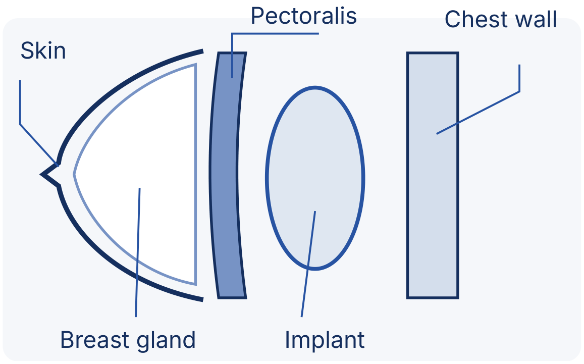
Schematics only, not to scale: not your actual surgical plan and not a guarantee of results.
Layered closure
Layered sutures close deeper tissues; sutures, skin adhesive and/or surgical tape close the skin. Scars are permanent, although they usually fade.
Early change, not the final result
Volume changes may be visible early. Swelling affects the appearance; shape and position settle over months. No precise size, symmetry or silhouette is guaranteed.


04 · After surgery and beyond
Recovery & lifelong monitoring
Healing is individual. Follow your written aftercare plan and continue monitoring both breast health and implants for the long term.
After surgery
Dressings and an individually prescribed support bra.
Early weeks
Soreness and swelling may last weeks; follow-up guides activity.
Next months
Swelling settles and scars mature at a variable pace.
Long term
Clinical review, screening and device-specific monitoring.
These are general stages, not deadlines or guaranteed recovery times.
Your written aftercare plan
Ask about wound care, prescribed medicines, bathing, stitches, follow-up and the type and duration of bra use. Drains are not routine but may be used if needed. Protect incisions from tension, rubbing and excessive movement. Return to work, exercise and driving only when your team clears you; do not use a fixed 48-hour rule.
Implants are not lifetime devices
Future surgery, removal or replacement may be needed. There is no automatic ten-year replacement rule. Aging, gravity, hormones, pregnancy and weight changes can alter results; breastfeeding is not guaranteed. Continue clinical follow-up: an annual examination alone cannot exclude silent rupture.
Silicone implant imaging: a reference schedule
For asymptomatic silicone implants, the FDA recommends ultrasound or MRI at 5–6 years after placement, then every 2–3 years. This is a US FDA reference schedule; agree the appropriate follow-up plan with your treating team. Agree an individual, device-specific and locally appropriate plan. Symptoms need earlier assessment: do not wait for the screening date. Uncertain ultrasound findings may require MRI.
Routine breast-cancer screening is separate and follows age and risk guidance. Tell the mammography team you have implants. Keep your device card, manufacturer/model, serial numbers, date and side of placement, and operation report.
When to seek help
Seek prompt assessment for persistent or late swelling, a lump, pain, hardening or new asymmetry. After surgery, contact your team for fever, redness or worsening pain. Chest pain or shortness of breath requires emergency care.
Safety reference: FDA, implant labeling and rupture-screening guidance (opens in a new tab).
05 · Benefits and trade-offs
Risks & informed consent
Decide whether the likely benefits are worth the risks for you. Complications are possible, not inevitable, and vary with health, technique and the exact device.
Surgical and tissue risks
- Anesthesia complications; bleeding or hematoma; infection.
- Temporary or permanent changes in nipple or breast sensation.
- Poor scarring, delayed wound healing or skin loss; asymmetry.
- Persistent pain; further or revision surgery.
Implant-related risks
- Malposition or displacement.
- Leakage or rupture.
- Capsular contracture: tightening of scar tissue around the implant, sometimes painful.
- Seroma: fluid collection.
- Visible or palpable wrinkling/rippling.
Rare cancers around the capsule
BIA-ALCL is a rare lymphoma associated with breast implants, usually arising in fluid or scar capsule around the implant; it is not ordinary breast cancer. Risk is higher with textured implants. Other rare cancers of the capsule have been reported, including squamous cell carcinoma (SCC) and other lymphomas. Persistent late swelling, a lump, pain or a new change needs prompt assessment, sometimes imaging and fluid or tissue tests.
Reported systemic symptoms
Some patients report fatigue, “brain fog,” joint pain and other symptoms, often called breast implant illness (BII). Their causes and relationship to implants are not fully understood. This does not establish that implants cause autoimmune disease, and it should not be dismissed as proof of no possible association. Discuss symptoms and evaluation with your clinician.
Device-specific informed consent
Review benefits, limitations, alternatives (including fat transfer, lift or no surgery) and the manufacturer’s leaflet and decision information. Ask about the exact device’s surface, rupture and contracture risks, future operations, monitoring and costs. Breastfeeding may or may not be possible. This list is not comprehensive; discuss risks specific to you.
Safety reference: FDA, risks and complications of breast implants (opens in a new tab).
06 · Planning ahead
Choosing your surgeon & cost
Choose an appropriately qualified, experienced plastic surgeon and a care pathway you trust, not simply the lowest quote.
What to look for
- Appropriate qualifications and authorization to practice; relevant surgical training.
- Experience with breast augmentation, the proposed technique and specific implant device or fat transfer.
- An appropriately approved and equipped facility, qualified anesthesia care and a clear emergency plan.
- Time to discuss your priorities, realistic outcomes, alternatives and risks without pressure.
- Written aftercare, accessible follow-up and a plan for complications or future procedures.
Questions for your surgeon
Why this device, size, incision and pocket for my anatomy? Would a lift, fat transfer or no surgery better meet my goals? What are the trade-offs for activity, scars, sensation and breastfeeding?
Who manages urgent concerns, including after hours? Where will I be treated if an unexpected complication occurs?
Ask for an itemized estimate
Fees vary with the operation, surgeon, device, facility and practice location. Request a written estimate and ask whether financing is available, without assuming it is offered.
- Surgeon’s fee
- Anesthesia
- Facility
- Medical tests
- Bra & medicines
Budget beyond the operation
Confirm implant/device fees, anesthesia, facility costs, tests, postoperative bra and medicines, follow-up and imaging. Ask who pays for possible future surgery, implant removal or replacement, and whether any device warranty actually covers those costs.
Confirm your own insurance
Cosmetic augmentation and related complications are often excluded. Check your policy and obtain an insurer’s confirmation of any proposed coverage; do not assume local coverage or reimbursement.
Take the written plan home, read the device leaflet and allow time for questions before deciding. A trusted surgeon should explain both the reasons to proceed and the reasons to reconsider.
Book your consultation
Let’s talk about your goals
- Phone +961 1 746345
- WhatsApp +9613307854
- Email psi@psi-lb.com
- Address 63 Souraty Street, Hamra, Nassif Bldg, 1st Floor, Beirut, Lebanon
This guide provides general educational information only. It does not replace an in-person assessment and individualized advice from a qualified plastic surgeon. It makes no promise or guarantee about results, safety, device lifespan or breastfeeding. A treatment plan and device-specific informed consent are required before surgery. Not all techniques or devices described are offered or locally available.