
Skin Cancer Removal
Understanding removal, repair and the care that follows.

01 · The basics
Understanding treatment
A skin cancer diagnosis can be unsettling. Surgery aims to remove the cancer; reconstruction aims to preserve function and appearance as far as possible. Cancer control comes first.
Your diagnosis guides the plan
Biopsy and tissue examination establish the diagnosis. Treatment depends on the cancer type, size, location, depth and risk. Appearance alone cannot establish whether a spot is cancer. Some cancers need radiation or other oncologic treatment according to pathology and stage.
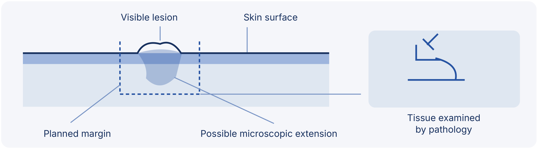
Illustrative cross-section, not to scale. Some lesions extend beyond what is visible; this is not the pattern of every cancer or an individual excision plan.
Conventional excision
The surgeon removes the lesion with a planned margin of surrounding tissue. The specimen is examined by pathology; results may arrive after surgery. If cancer remains at an examined edge, further excision may be needed. Some cases use intraoperative frozen-section evaluation.
Mohs is a distinct technique
For selected cancers, an appropriately trained Mohs surgeon removes mapped layers in stages and examines peripheral and deep margins, repeating as needed while the patient waits. Referral may be appropriate. It is not required for every skin cancer.
Mohs and other frozen-section margin assessments are not interchangeable.
What does a clear margin mean?
No cancer is seen at the examined tissue edges. This is not a guarantee against recurrence or distant spread, and it is not a promise of cure.
02 · Before surgery
Consultation & preparation
An open discussion helps the team choose a safe, appropriate plan. Share your concerns about both cancer treatment and the effect on your appearance.
Bring and discuss
- Biopsy/pathology reports, the site of the lesion and previous skin cancer treatments.
- Medical conditions, allergies, previous operations and any healing problems.
- All medicines, vitamins and herbal supplements; alcohol, tobacco and other substances.
- Your priorities, support at home and questions about scars, function and further treatment.
Your surgeon will review
- Your health, risk factors, skin and the biopsy findings; photographs may document the site.
- Removal options, margin assessment and whether a Mohs referral is appropriate.
- Repair choices, immediate or delayed reconstruction, anesthesia and the place of care.
- Expected benefits, limitations, complications and the follow-up plan.
Personalized preparation
- Medical evaluation
- Medicine review
- Smoking advice
- Transport & support
Tests or medical assessment may be requested. Follow the team’s instructions on smoking and any medicine adjustments, including aspirin, anti-inflammatory drugs and supplements. Do not stop prescribed medicines without your surgeon’s directions.
Plan the practical details
Confirm where treatment will take place and any fasting instructions. If sedation or general anesthesia is planned, arrange transport and an adult to stay with you for at least the first night, as instructed. Ask who will communicate pathology results and when to return.
Ask questions before signing consent. Understanding the proposed removal and repair matters more than rushing a decision.
03 · On the day
Removal & reconstruction
The operation and repair are tailored to the lesion and the margin strategy. Reconstruction may be immediate or delayed while tissue results or further excision are awaited.
Anesthesia
Local anesthesia, intravenous sedation or general anesthesia may be used. The team recommends the option suited to the extent of surgery and your health.
Removal & margin assessment
The lesion is removed by the selected method. Conventional excision, intraoperative frozen-section evaluation and specialized Mohs surgery are different approaches; the plan determines how and when margins are assessed.
Repair of the defect
Small defects may close directly with stitches. Larger or more complex wounds may need a local flap or skin graft. The choice depends on location, depth, available tissue and cancer clearance.
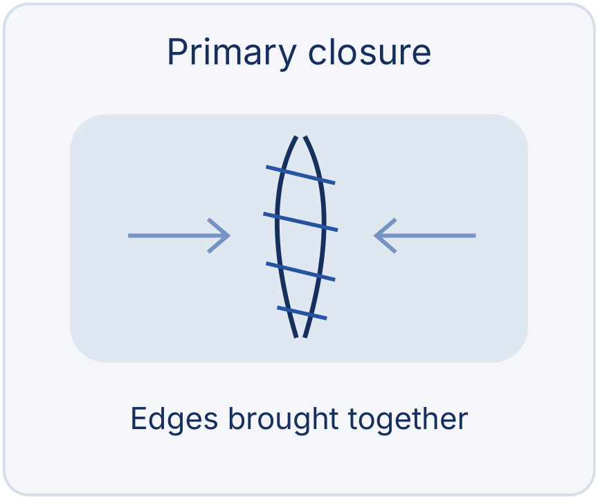
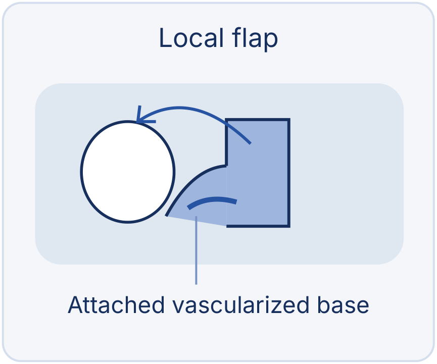
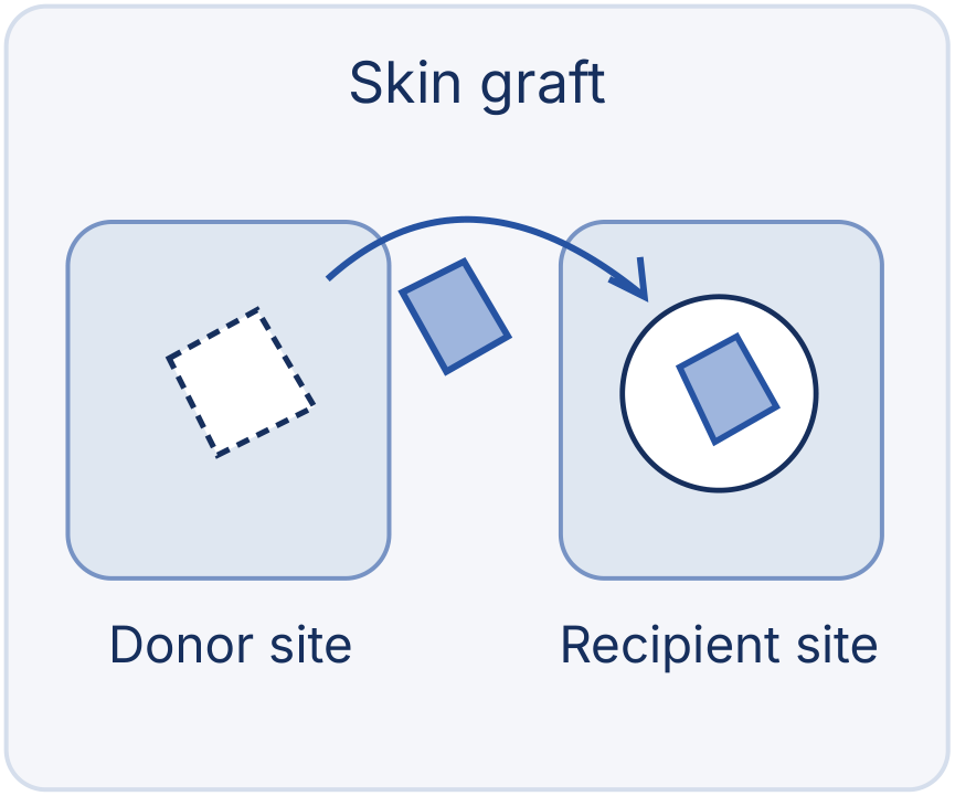
Schematics, not to scale. A flap moves nearby skin while retaining an attached blood supply. A graft is a separate skin patch transferred from a donor site, without its original blood supply connection; it develops a supply from the recipient wound. The donor site is also a wound.
Dressing & instructions
A dressing or bandage protects the site. You receive wound-care advice, a plan for results and follow-up, and instructions for any donor-site wound.
Suture lines may follow natural creases where feasible, but every operation leaves scars. No reconstruction can guarantee a normal appearance or function.
04 · After surgery
Recovery, results & follow-up
Healing is individual. Follow the cleaning, dressing and topical or oral medicine instructions exactly as prescribed; there is no single timetable for every repair.
Early protection
Some tenderness, redness or a small amount of drainage may occur. Protect the wound and any donor site.
Wound review
Dressings, stitches, light activity and return to exercise are reviewed according to your operation and healing.
Scar maturation
Scars change over months and may take a year or longer to soften and fade. They do not disappear.
Long-term care
Continue skin checks, sun protection and the dermatologist follow-up schedule tailored to your risk.
Stages of care, not fixed day-by-day deadlines.
Protect the repair
- Keep the site clean and protected as instructed; use only the cleaning products and topical medicine prescribed.
- Avoid tension, abrasion and excessive movement that stress the wound or stitches. Ask when to wash, change dressings, resume activities and return.
- Protect healthy skin and the healing site from sun exposure according to advice. Do not put sunscreen on an open wound.
When to contact your team
Contact the treating team for persistent or increasing bleeding, redness, heat or pain, or other concerning changes. Ask for a clear contact plan before leaving.
Results and continued surveillance
Permanent scars, color or texture differences and, sometimes, disfigurement can remain. Staged refinements may be needed. Review your pathology results and any recommendation for further excision, radiation or other treatment. Examine your skin regularly; report a new or changing lesion sooner rather than waiting for the next visit. Dermatology reviews are individualized and may be more frequent than yearly.
05 · An informed decision
Risks & safety
Discuss the expected benefits, limitations and your individual risks before consent. Surgery and reconstruction have risks even when carefully planned.
Allergic reactions
- Reactions to tapes, sutures or glues.
- Reactions to blood products, topical preparations or injected agents.
Surgery & anesthesia
- Anesthesia complications.
- Excessive bleeding.
- Infection.
Nerves & sensation
- Changed skin sensation, which may persist.
- Temporary or permanent injury to nerves, blood vessels or muscles.
Healing & reconstruction
- Poor wound healing and permanent or unfavorable scars.
- A graft may fail to take; a flap may have circulation problems.
- Further surgery or revision may be necessary.
Disease risks: separate from surgical complications
Cancer may recur locally or spread to distant sites, depending on its type and stage. These are risks of the disease, not a claim that surgery causes spread. Clear examined margins do not eliminate these risks. Your team will explain what applies to your diagnosis.
Make the safety plan clear
- Which risks are most important in my case, and how will they be reduced?
- What happens if margins are involved, a graft fails or healing is delayed?
- Who coordinates pathology, further treatment and skin surveillance?
- How do I contact the team if recovery is not going as expected?
No treatment can promise a perfect repair or freedom from recurrence. Ask about alternatives and any uncertainty specific to your case.
06 · Planning ahead
Costs & choosing your surgeon
Ask for an individualized written estimate. Costs depend on the extent of removal, the repair, anesthesia and the place of care; no universal price applies.
- Surgeon’s fee
- Anesthesia
- Clinic or hospital
- Tests & pathology
- Medicines & dressings
Confirm whether reconstruction, donor-site care, follow-up and any additional excision or staged procedure are included, and what could be billed separately.
Check insurance before surgery
Your plan may cover medically necessary excision and reconstruction. Confirm coverage, prior authorization, limits and required documents with your insurer before surgery. Do not assume reimbursement is guaranteed.
Choose experience and clear communication
- A qualified surgeon experienced in skin-cancer excision and reconstruction.
- A facility appropriate for your procedure, anesthesia and medical needs.
- A clear explanation of removal, margin assessment, repair, risks and realistic outcomes.
- Coordination with dermatology, pathology and oncology when needed, including referral for specialized techniques.
- A written aftercare and follow-up plan, with a reliable contact route.
Experience, continuity of care and confidence in the treatment plan matter alongside cost.
07 · Your next step
Discuss your treatment plan
- Phone +961 1 746345
- WhatsApp +9613307854
- Email psi@psi-lb.com
- Address 63 Souraty Street, Hamra, Nassif Bldg, 1st Floor, Beirut, Lebanon
This guide is for general educational purposes only. It makes no promise or guarantee about the outcome of any procedure and is not a substitute for a thorough, in-person consultation with a qualified plastic surgeon.